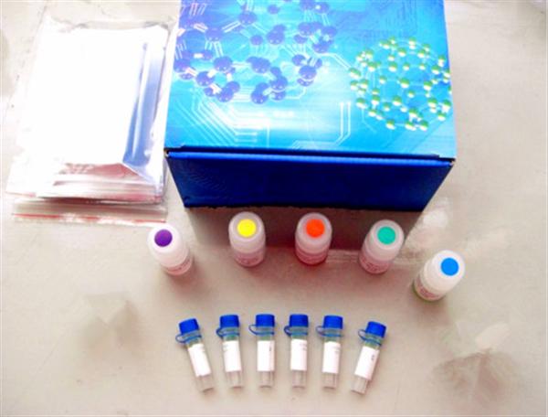
The most common method for detecting antigens in the elisa kit:
The steps are as follows:
1) A specific antibody is linked to a solid phase carrier to form a solid phase antibody. Washing removes unbound antibodies and impurities.
2) Add the specimen to be inspected and keep warm. The antigen in the specimen binds to the solid phase antibody to form a solid phase antigen-antibody complex. Wash to remove other unbound material.
3) Add the enzyme-labeled antibody and keep the reaction. The antigen on the solid phase immune complex binds to the enzyme-labeled antibody. Unbound enzyme-labeled antibody was thoroughly washed. The amount of enzyme carried on the solid support at this time is related to the amount of the antigen to be tested in the specimen.
4) Add substrate to develop color. The enzyme on the solid phase catalyzes the substrate to become a colored product. The amount of antigen in the specimen is measured by colorimetry.

In clinical tests, this method is suitable for testing macromolecular antigens such as various proteins such as HBsAg, HBeAg, AFP, hCG, and the like. As long as the heterologous antibody against the test antigen is obtained, the method can be established by coating the solid phase carrier and preparing the enzyme conjugate. For example, if the source of the antibody is an antiserum, the antibodies for coating and enzymatic labeling are preferably taken from animals of different species. If monoclonal antibodies are used, two monoclonal antibodies directed against different determinants of the antigen are typically selected for coating the solid support and preparing the enzyme conjugate, respectively. The two-site sandwich method has high specificity, and the test specimen and the enzyme-labeled antibody can be incubated together for one-step detection.
In the one-step assay, when the content of the test antigen in the specimen is high, the excess antigen is combined with the solid phase antibody and the enzyme-labeled antibody, respectively, and no "sandwich complex" is formed. Similar to the post-band phenomenon of excess antigen in the precipitation reaction, the absorbance of the color developed after the reaction (located on the excess of the antigen) is the same as the absorbance of the standard curve (located on the excess band of the antibody) at an antigen concentration (see 1.3). .2, Figure 1-4), if measured according to the usual method, the result will be lower than the actual content. This phenomenon is called the hook effect, because the standard curve is hooked and bent after reaching the peak. . When the hook effect is severe, the reaction may even show no false negative results. Therefore, when using a one-step reagent to measure substances with abnormally high levels of specimens (such as HBsAg, AFP, and urine hCG in serum), the highest value of the measurable range should be noted. Preparation of such agents with high affinity monoclonal antibodies can attenuate the hook effect.
If a plurality of identical determinants, such as the a determinant of HBsAg, are present at different sites of the molecule being tested, the same monoclonal antibody for this determination can also be used to coat the solid phase and prepare the enzyme conjugate, respectively. However, in the detection of HBsAg, attention should be paid to the subtype problem. HBsAg has four subtypes of adr, adw, ayr, and ayw. Although each subtype has the same reactivity of a determinant, this is also the use of monoclonal antibody as a sandwich method. Attention to the problem. The double antibody sandwich method is suitable for the determination of macromolecular antigens of divalent or higher valence, but is not suitable for the determination of hapten and small molecule monovalent antigens because it cannot form a two-point sandwich.
Smart Uv Printer,3D Photo Printer,3D Emboss Back Skin Printer,3D Relief Back Film Printer
Shenzhen TUOLI Electronic Technology Co., Ltd. , https://www.szhydrogelprotector.com